
Pregnancy
When Does a Baby's Heartbeat Appear or Become Audible?
Fetal heart activity is sometimes visible around the 6th week on an early ultrasound. Why are the examination method, embryo size, and timing of follow-up important?

Most Frequently Asked Question: When Is a Heartbeat Detected?
A baby's cardiac activity can sometimes be seen on transvaginal ultrasound around the 6th week of pregnancy. This is not a deadline or a definitive rule for every pregnancy. How early the examination is performed, the method used, and the size of the embryo affect the result.
Seeing and hearing are separate concepts. In an early ultrasound, the specialist assesses the movement of cardiac tissue within the embryo. Listening to the sound with a handheld Doppler is another method and is usually used later; listening to the sound at the first appointment is not mandatory.
The absence of cardiac activity at the first examination alone is not a diagnosis of pregnancy loss. An earlier stage or a developmental issue are possible reasons. The doctor combines measurements and previous data, and if necessary, schedules a repeat ultrasound after an appropriate interval.
How Does Early Heart Activity Appear on an Ultrasound Scan?
In the 6th week of pregnancy, the embryo is very small, about the size of a pea or a lentil (approximately 6 mm). At this stage, it is covered with transparent skin, and its body parts are just beginning to form. During an ultrasound examination, the specialist looks for the embryo (fetal pole) inside the gestational sac.
Cardiac activity appears as a rapidly flickering dot within this tiny embryo. This is an image created by sound waves reflecting off the moving cardiac tissue. When your doctor or sonographer sees this flicker, it indicates the presence of cardiac activity at that moment of the examination and can be reassuring information. However, remember that the results of an early examination may not be 100% accurate, and sometimes additional examinations are needed to confirm the diagnosis.
Last Menstrual Period (LMP) and Late Ovulation Factor
The week of pregnancy is usually calculated from the first day of the last menstrual period (LMP). However, this calculation method is not always accurate. International medical guidelines, including recommendations from the National Institute for Health and Care Excellence (NICE) in the UK, advise against making decisions about when a heartbeat should be seen based solely on LMP.
This is because the duration of the menstrual cycle varies from woman to woman. In some women, ovulation may occur later in the cycle, not in the middle. This means that fertilization and pregnancy started later than expected. As a result, a pregnancy you consider 6-7 weeks based on LMP may actually be 5 weeks. This one-week difference is a sufficient reason for cardiac activity not yet being visible. This is precisely why doctors rely more on ultrasound measurements (CRL) for diagnosis.
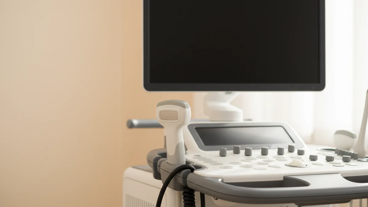
Vaginal vs. Abdominal Ultrasound: Differences in Methods
Transvaginal ultrasound is performed with a vaginal probe and can provide a clearer image in early pregnancy. Transabdominal ultrasound is performed over the abdomen. Since the imaging capabilities of these two methods are different, the result and repeat examination plan may vary in the same week.
NICE recommends offering vaginal ultrasound to assess the location and development of an early pregnancy. If this method is not suitable for you or you do not wish it, an abdominal examination may be offered, and its limitations explained. It is possible to ask questions about the examination and clarify the issue of consent.
When comparing examinations, clarify the method. Do not enlarge the image on the phone and decide for yourself about the heartbeat or measurement; the report and the specialist's interpretation should be considered together.
CRL Measurement and Repeat Scan: What to Expect?
If the embryo is visible but cardiac activity is not, the doctor assesses the size of the embryo. If the embryo itself is not visible, separate criteria apply to the size of the sac. This measurement is called CRL (Crown-Rump Length), which means "length from crown to rump." This is an important measurement used in assessing early pregnancy duration. For accurate diagnosis, there are waiting periods and criteria established by organizations like NICE. This is done to minimize the risk of misdiagnosis.
The table below shows NICE's pre-diagnosis approach when cardiac activity is not detected in a visible embryo. These are not thresholds for self-diagnosis at home; sometimes additional examination is needed. All minimum intervals are calculated from the first examination:
| ultrasound Type | CRL Measurement | Cardiac Activity | Next Step |
|---|---|---|---|
| Transvaginal ultrasound | Less than 7.0 mm | Not visible | Repeat examination scheduled after a minimum of 7 days. |
| Transvaginal ultrasound | 7.0 mm or larger | Not visible | A second specialist's opinion is sought and/or a repeat examination is scheduled after a minimum of 7 days. |
| Transabdominal ultrasound | Any size | Not visible | CRL measurement is noted, and a repeat examination is scheduled after a minimum of 14 days. |
This waiting period has no negative impact on the outcome of the pregnancy. It is simply a safety measure to avoid making a hasty and potentially incorrect decision. If you experience any new or worsening symptoms during this period, you should contact your doctor immediately.
Listening to the Heartbeat: Doppler Devices and Risks of Home Use
A handheld Doppler may be used by a doctor or midwife in later pregnancy appointments. When the sound is detected with it depends on the method, experience, and individual situation; a fixed week cannot be determined for everyone. This is a very exciting moment. However, there are serious warnings regarding home Doppler devices recently on sale.
Using a home Doppler device is not recommended. There are several important reasons for this:
- Misleading sense of security: Hearing a heartbeat does not mean the baby is completely healthy. For example, a baby whose movements are decreasing but still has a heartbeat may need emergency care. Delaying a visit to the doctor by hearing a heartbeat and thinking "everything is fine" can be dangerous.
- Risk of misinterpretation: An inexperienced person can easily mistake the baby's heartbeat for the mother's own pulse or the sound of blood flow from the placenta.
- Unnecessary anxiety: Not being able to find the heartbeat due to the baby's position or early pregnancy can cause great stress and anxiety, even if there is no problem.
Entrusting the assessment of your baby's health to professional medical staff is the safest way.
Does One Heartbeat Guarantee Health?
Seeing cardiac activity on an early ultrasound can be reassuring news: the movement of cardiac tissue was assessed at that moment of the examination. However, seeing cardiac activity once is not a guarantee of problem-free development throughout the entire pregnancy.
Planned monitoring answers different questions at various stages of pregnancy. Do not cancel subsequent examinations just because the initial result was good. If new bleeding or pain occurs, previous cardiac activity is not a reason to disregard that symptom.
You can ask your doctor what the result currently indicates, what questions remain open, and the purpose of the next appointment. The visibility or speed of the heartbeat is also not a method for determining the baby's gender.
When to Seek Emergency Medical Help?
Seek medical advice on the same day if you have new spotting or vaginal bleeding, or persistent abdominal and pelvic pain. Bleeding does not always mean a loss, but the cause may need to be clarified.
Seek emergency care immediately if you have severe or sudden abdominal pain, especially one-sided pain; shoulder tip pain; loss of consciousness or severe dizziness; heavy bleeding and rapidly worsening condition. Do not wait for a certain number of pads to be filled or for the scheduled ultrasound day. Severe symptoms are important even without visible bleeding.
In later pregnancy, decreased movement or a change in the usual pattern also requires immediate contact with maternity services. Hearing a heartbeat at home does not replace this consultation.
Preparing for a Doctor's Appointment: What Questions to Ask?
It's normal to feel anxious during a doctor's appointment and forget to ask some points. Writing down your questions before the appointment can help you. Here are some useful questions you can discuss with your doctor:
- “What do the results of this ultrasound specifically mean?”
- “Does the embryo's size (CRL) match the expected week according to my last menstrual period?”
- “If cardiac activity was not seen, what is our next step and when is the repeat examination scheduled?”
- “What symptoms should I pay special attention to during the waiting period, and in what case should I contact you immediately?”
- “What do these results mean for the course of my pregnancy?”
Your doctor should understand your concerns and answer all your questions clearly. Remember, this is your body and your baby. Being informed will help you get through this sensitive period more calmly.

Conclusion: Patience and Professional Care
Seeing or hearing your baby's heartbeat for the first time is an unforgettable moment for every parent. However, a slight delay in this moment is not always a sign of a problem. The first weeks of pregnancy can be full of uncertainties. Most importantly, be patient, follow your doctor's instructions, and be attentive to your body's signals. Avoid drawing hasty conclusions based on the result of a single early examination. Accurate diagnosis sometimes requires time and repeat examinations.
You can use Anacan's Pregnancy Calendar to track your doctor's appointments and examination dates. We wish you a healthy and comfortable pregnancy!
Frequently Asked Questions
At what week is a baby's heartbeat visible at the earliest?
Heart activity can sometimes be visually seen on a transvaginal ultrasound around the 6th week of pregnancy. However, this is not true for everyone and may not be visible due to the pregnancy being at an earlier stage.
If no heartbeat is seen on the first ultrasound, does this mean miscarriage?
No, this cannot be said from a single result. The doctor evaluates the examination method, the size of the visible embryo, and historical data together. An appropriate minimum interval for follow-up and sometimes additional examination is needed; do not wait for that date if new severe symptoms arise.
Are home Doppler devices safe to use?
No, medical professionals do not recommend the use of home Doppler devices. These devices can create a misleading sense of security or cause unnecessary anxiety. The assessment of the baby's health should be performed by professionals.
When is the sound of the heartbeat audible?
Listening with a handheld Doppler is usually possible at later appointments, but there is no fixed week for everyone. It is not the same method as assessing visual heart activity on an early ultrasound. Not hearing the sound at the first appointment is not a diagnosis on its own.
Is waiting for a repeat ultrasound scan harmful to the baby?
According to NICE, waiting for a planned diagnostic repeat ultrasound does not harm the outcome of the pregnancy. The minimum interval is chosen based on the examination method and measurement. However, if new bleeding, pain, or general worsening occurs, seek help without waiting for that date.
What is CRL and why is it so important?
CRL (Crown-Rump Length) is the length of the embryo from its crown to its rump. This is the most reliable measurement in the first trimester that allows for more accurate dating of the pregnancy than the last menstrual period. Doctors rely on this measurement to determine if heart activity is present at the expected time.